
BATTENKILL VETERINARY
516 State Route 29 (518) 692-2227
Greenwich, NY 12834 battenkillvet4u@yahoo.com
Medical Conditions and Images
Click on the disease listed by the picture for more information. Please be advised that some of the following photographs are graphic and may not be suitable for some people.

This poor pitbull tangled with a porcupine and didn't come out on the winning end.
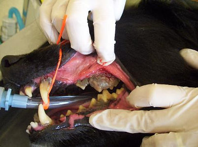
Below is an example of severe dental disease right before and after an ultrasonic dental cleaning (prior to extractions). The severe root exposure was not completely seen when the teeth were covered in tartar. These teeth were not stable and were removed (extracted) after this photo was taken.
Dental Disease


Lipoma
The above images are before and after surgical removal of a lipoma. Lipomas are tumors composed of adipose tissue and are also known as fatty tumors. Almost all lipomas are benign and slow-growing. In this case, the lipoma was interfering with the patient's ability to walk and therefore it was removed.

Splenomegaly
Splenomegaly or enlargement of the spleen can occur for several reasons. In this case, the patient had malignant cancer that had spread to the spleen. The spleen in this image is approximately 10 times larger than the average spleen.


Pyometra
This is a uterus that is full of pus (pyometra). This female dog was not spayed when younger and developed a life-threatening infection in her uterus. The treatment for a pyometra is the spay surgery (ovariohysterectomy) but it is high risk and some dogs do not survive the procedure. The spay surgery is less risky when performed on a young dog, preferably before she has a heat cycle or puppies. This can also happen in cats that are not spayed.
Bladder Stones
Bladder stones (uroliths) may form due to diet, genetic predispositions, or infections. They can cause significant disease if left untreated by plugging the outflow of urine. These bladder stones were surgically removed.
Intestinal Parasites - Whipworms
This is a microscopic view of whipworm eggs. Each football shaped object in the image is an egg. Whipworms can cause severe disease if not appropriately treated. Annual fecal examinations allow veterinarians to check for intestinal parasites.
Vulvoplasty, pictured below, is a procedure performed to remove excess fat and skin that covers or 'hoods' the vulva. Dogs with a hooded vulva are predisposed to urinary tract and skin infections and may benefit from surgical correction.
Vulvoplasty


Gastrointestinal Foreign Body
You've all heard the story of a dog or cat eating something they should not - shoes, furniture, toys. Gastrointestinal foreign bodies often result in emergency surgery as the objects can obstruct intestinal flow and cause significant damage. The upper left image is carpet removed from a dog's intestine. The image to the left is of a cat that ate string (linear foreign body). Above is a kitten that had eaten a coiled plastic key ring.
Sarcoptes
Sarcoptes is the culprit behind sarcoptic mange. This mite burrows in the skin and causes extreme itchiness. This image was taken through a microscope and the two round critters are the sarcoptes mites.
Bite Wounds
This is an example of a cat with a bite wound. If left untreated, these often become infected and are unable to heal.